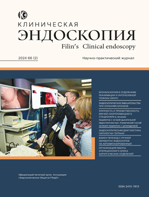
Scientific and Practical Peer-Reviewed Medical Journal for Specialists in Radiology, Endoscopy, Surgery, and Other Related Fields. The journal is dedicated to scientific issues in endoscopy and radiology, including topics in experimental and clinical medicine, scientific reviews and lectures for practicing physicians, case studies from clinical practice, as well as information about the latest scientific forums in Russia and abroad on major issues in radiology and endoscopy.
Current issue
Vol 67, No 3 (2025)
LEADING ARTICLE
5-9 143
Abstract
The paper presents an impersonal forensic medical examination of a case of colon perforation after endoscopic submucosal dissection of cecum epitelial neoplasm.
ADMINISTRATIVE ISSUES
10-17 188
Abstract
Infections pose a serious problem in ensuring epidemiological safety during endoscopic interventions. The experience of the last two decades shows that changes in pathogens, with ESCAPE pathogens dominating, complicate control of outbreaks, increasing the number of affected patients and mortality rates. Unfortunately, in our country there is virtually no registration of infections associated with endoscopic interventions. The aim of this work is to summarize the world literature data on the prevalence and characteristics of infections associated with diagnostic and therapeutic endoscopic interventions. Materials and methods. We conducted a systematic review of 47 literature sources for the period from 1991 to 2025 from the PubMed and Embase databases to study the distribution of cases/outbreaks of HAI in endoscopy with an analysis of the causes of their occurrence and the most effective strategies for their relief. Results. The article presents data on the prevalence of HAI in therapeutic and diagnostic endoscopy. The features of the endogenous type of infection are analyzed, which is mainly determined by internal risk factors on the part of the patient, the complexity of the intervention, the competencies of the doctor and the effectiveness of antibiotic prophylaxis, if used. The leading risk factors for outbreaks in diagnostic endoscopy are analyzed. Discussions continued the effectiveness of high-level disinfection and the acceptability of endoscope sterilization, as well as the need to improve the effectiveness of microbiological quality control of endoscope processing. The place of the Epidemiological Safety System (ESS) of endoscopic interventions, developed in our country, in a set of measures to prevent HAI was determined.
18-22 226
Abstract
The article examines the issues of regulatory support for the activities of the endoscopic service, the quality and safety of medical activities.
M. S. Burdyukov,
I. E. Usyatinskaya,
A. V. Alekseev,
R. O. Kuvaev,
M. P. Korolev,
N. A. Aliev,
I. I. Korzheva,
M. N. Kuzin,
S. P. Petrov,
V. V. Amirova,
V. V. Veselov,
S. V. Kashin,
D. V. Gusev
23-35 165
Abstract
The Federal Law No. 323-FZ of November 21, 2011 establishes the requirements for the content and format of the “Informed Voluntary Consent” (IVC) form for medical interventions. This document serves as a legal guarantee of protecting the rights of both patients and healthcare professionals. The article discusses the procedure of signing the IVC and highlights its key sections that require special consideration. The most important are the sections “Indications” and “Contraindications for medical intervention.” Indications provide patients with information about the necessity of a particular procedure and the possible consequences of refusal, ensuring awareness and informed decision-making. The section on contraindications reflects potential health and life risks related to the patient’s condition or to the specifics of the intervention, including possible complications. The patient’s signature confirms awareness and consent, while also providing legal protection for the healthcare institution and staff in case of adverse outcomes or legal disputes. Special attention is paid to the section “Risks associated with medical care.” The list of potential adverse events (for example, during colonoscopy) can be extensive, which makes it impossible to include every detail. Therefore, the authors propose standardization and adaptation of such risks for inclusion in the IVC form. This approach allows achieving a balance between comprehensive patient information and practical usability of the document.
36-40 256
Abstract
The purpose of the article is to introduce readers to the basics of ultrasound and the main principles of its operation in order to effectively manage variables that can be classified as operator-dependent and collect the most complete information from each study, avoiding pitfalls and errors in diagnosis.
CLINICAL OBSERVATIONS
41-49 180
Abstract
Respiratory tract pathology occupies a leading place in the structure of morbidity. The high prevalence of chronic bronchitis, bronchial asthma and chronic obstructive pulmonary disease justify the need to study, interpret and analyze the endoscopic semiotics of this pathology, correctly classify the identified inflammatory changes in the bronchi. The article discusses the classification of bronchitis, clinical observations.
S. V. Dzhantukhanova,
Yu. G. Starkov,
M. E. Timofeev,
A. I. Vagapov,
O. T. Imaraliev,
O. B. Abu-Haidar
50-55 110
Abstract
Objective of the study. To present the experience of successfully treating a patient with gastric stalk wall suture failure after proximal gastrectomy with lower thoracic esophagectomy using an endoluminal suture device. Clinical case. A 54-year-old patient with a malignant neoplasm of the proximal stomach with extension to the lower thoracic esophagus (cT3N1M0, stage III) was treated at the Oncology Center from July to October 2025. The patient underwent proximal subtotal gastrectomy with resection of the distal stomach. Postoperatively, the patient developed gastric stalk suture failure, requiring pleural drainage and repeat surgery. Results. Endoscopic suturing of the gastric stalk wall defect using the Apollo OverStitch endoluminal suture device resulted in a positive clinical outcome in the treatment of a complex postoperative complication. It should be noted that the patient was restored to oral nutrition within two weeks. Conclusion. Our data demonstrate the high potential of the Apollo Overstitch™ endoluminal suturing device for endoscopic closure of gastric stalk wall defects caused by suture failure.
GASTROENTEROLOGY
56-66 134
Abstract
Objective. To present current perspectives on the epidemiology, etiology, and pathogenesis of eosinophilic esophagitis (EoE). Main Points. Eosinophilic esophagitis is a chronic, inflammatory, immune-mediated disease of the esophagus. Its pathogenesis is based on a predisposition to mount an immune response via the activation of type 2 T-helper (Th2) cells, which, upon contact with exogenous allergens, express a highly active group of cytokines. The high concentration of Th2 cytokines in the esophageal mucosa leads to eosinophilic infiltration throughout the entire length of the esophageal wall (from the proximal to the distal segments), the development of chronic inflammation within the epithelium and the lamina propria, and the development of subepithelial fibrosis. Patients with EoE often have concomitant Th2-associated inflammatory diseases (such as bronchial asthma, rhinitis, conjunctivitis, hay fever, etc.). Timely diagnosis and adequate treatment of eosinophilic esophagitis can prevent the development of strictures and other complications of the disease.
M. A. Paronyan,
S. S. Pirogov,
V. I. Ryabtseva,
G. F. Minibaeva,
D. G. Sukhin,
V. M. Khomyakov,
I. V. Kolobaev,
A. B. Ryabov,
V. S. Surkova,
A. D. Kaprin
67-73 460
Abstract
Introduction: Gastric cancer in young patients is of particular interest due to a number of clinical and endoscopic features. Despite of the general downward trend in gastric cancer incidence, in recent years there has been a steady increase in cases among young patients, which determines the relevance of studying the characteristics of this pathology. Objective: Evaluation of the results of endoscopic diagnostics of cases of gastric cancer detected in patients under 30 years of age in the endoscopy department of the P.A. Hertsen Moscow Oncology Research Institute. Materials and methods: A retrospective analysis of 27 cases of gastric cancer in patients under 30 years of age examined at the P.A. Hertsen Moscow Oncology Research Institute in the period 2012-2024 was conducted. Expert-class endoscopic equipment with near-focus endoscopy technology (NBI Near Focus) was used. Results: Infiltrative forms were observed in 100% of cases (70% - type III according to Borrmann classification, 30% - type IV). Total gastric involvement was detected in 41% of cases, with extension to the esophagus and/or duodenum in 45% of them. 60% of cases were diagnosed at stage IV. Histologically - poorly differentiated adenocarcinoma with a signet ring cell component predominated (89%). A case of association with Lynch syndrome was identified. Conclusion: Gastric cancer in young patients is characterized by an aggressive course and a predominance of infiltrative forms, which requires the development of special algorithms for early diagnosis and increased oncological alertness.
COLOPROCTOLOGY
74-85 169
Abstract
The aim of the study was to substantiate the endoscopic diagnosis of chronic asymptomatic colitis (CAC). Material and methods. A continuous retrospective cross-sectional study. 4099 videocolonoscopy protocols and histologic examination results for 2022-2024 were studied. We took into account the fact of conclusion of CAC in the study protocol, localization of CAC, subjective endoscopic features/criteria like color, surface and structure of mucosa, density and regularity of vascular pattern, presence and localization of diverticula, mucus, intestinal tone, presence and types of neoplasms. A database including passport information and patient complaints was created. Additionally, we used microscopic evaluation of biopsy specimens at the conclusion of “CAC”, which were stained using different histochemical techniques depending on the purpose of the analysis. Morphometric analysis included quantification of total mucosal thickness, thickness of the covering epithelium, thickness of the basal membrane, area of fibrosis in the proper lamina, cytoplasmic area of bocaloid cells of the covering epithelium, and number of inflammatory cells per crypt. Statistical analysis was performed using the licensed package of applied programs IBM SPSS Statistics-22. Results. The total frequency of CAC findings in the videocolonoscopy protocol amounted to 40% (1641 patients) and was associated with the patient’s age (χ2=162,219, p=0,0000). The prevalence of mucosal changes in all parts of the colon in CAC was 58% (956 patients). The endoscopic sign of CAC was thickening of the vascular pattern (39%,1614 cases) and traces of mucus after lavage preparation (350 observations). Diverticula were described in 21% of findings (872 cases). Morphometry revealed significant differences between normal, CAC and diverticulosis in terms of changes in mucosal thickness (χ2= 10.96486, p =0.00437), cover epithelium thickness (χ2= 13, 16667, p =0.00138), basal membrane thickness (χ2= 21,33333, p =0.00002), area of bocaloid cells (χ2= 11,20000, p =0.00370), and area of glandular epithelium (χ2= 6,88888, p =0.03192). Conclusions. Chronic asymptomatic colitis is an important diagnostic finding detected by morphologic examination of the colonic mucosa. Differentiation of CAC from drug-induced lesions and early forms of inflammatory bowel disease requires a comprehensive evaluation, including clinical, endoscopic, morphologic, and anamnestic data. Taking into account the potential association of CAC with neoplasia, it is advisable to form a unified approach to diagnosis, verification, endoscopic report and follow-up of patients with this pathology.
M. M. Lokhmatov,
G. A. Korolev,
E. I. Khvatova,
A. V. Tupylenko,
V. I. Oldakovskiy,
T. N. Budkina,
E. Yu. Dyakonova
86-92 149
Abstract
Relevance. Familial adenomatous polyposis syndrome is a severe6 cancer-associated disease that usually manifests itself in childhood. If left untreated, it leads to the development of colorectal cancer in 100%. The purpose of this study is to provide up-to-date information on the etiology, pathogenesis, diagnosis, and treatment of patients with familial adenomatous polyposis. The main part. Familial adenomatous polyposis is a genetically determined disease that develops as a result of a mutation in the APC suppressor gene. The vast majority of adenomatous polyps are formed in the colon. An adenoma is a neoplastic growth that can become an adenocarcinoma. However, due to the fact that there can be several thousand polyps in the intestinal lumen in FAP, the risk of malignancy is close to 100%. Conclusion. The only radical treatment for SAP is total colectomy. However, adenomas can also form in the small intestine, requiring ongoing monitoring and endoscopic treatment, as duodenal cancer is the second leading cause of death in patients with SAP.
NURSING
93-100 114
Abstract
Goal: to demonstrate the necessity and importance of maintaining specialized medical documentation in the endoscopy department to ensure the safety of patients and medical staff, reduce the risk of healthcare-associated infections (HAIs), control the quality of medical services, optimize resources, and comply with the regulatory requirements of the Russian Federation legislation. To examine how systematic and accurate record-keeping contributes to improving the efficiency of the department’s operations and reducing the risk of issues during inspections by regulatory authorities. Materials and methods: the study is based on an analysis of the practice of maintaining medical documentation in the endoscopy department of a medical organization. Various types of registration logs (for procedures, disinfection, consumables accounting, staff training, etc.) and their functional purposes were reviewed. The research methods included systematization of regulatory requirements, comparative analysis of the content of logs, and assessment of their impact on enhancing the safety and quality of medical care.
INFORMATION
ISSN 2415-7813 (Print)


